|
The SSM uses a
very sensitive magnetic field detector called a
SQUID (Superconducting Quantum Interference Device).
This device is made using superconductors that must
be cooled to cryogenic temperatures (below 80K or
about -200 degrees Celsius).
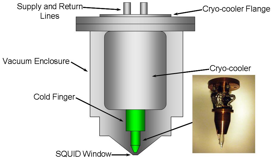
The SQUID is mounted
to the end of a sapphire rod (clear, cone-shaped rod
at the bottom of the lower right picture below).
Because it must be cooled to very low temperatures
(less than the temperature where oxygen liquifies),
the cooled components must be contained in a vacuum
chamber. A thin diamond window separates the
SQUID from the room-temperature sample being
scanned.
The SQUID can be seen
slightly to the right of center in the picture
below. The square opening in the center of the
SQUID is about 30 microns across, smaller than the
diameter of a human hair. You would see this
view of the SQUID if you aimed a microscope straight
up from beneath the cold finger assembly shown
above.

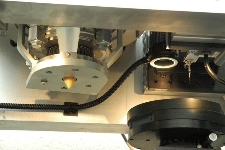
Here is a close-up
showing the window and vacuum assembly (the window
is at the bottom of the brown cone). The top
of the scanning stages can be seen to the lower
right.
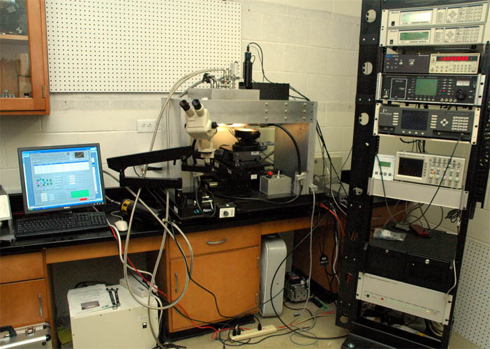
A computer controls
an X-Y-Z stage that scans the sample under the
SQUID.
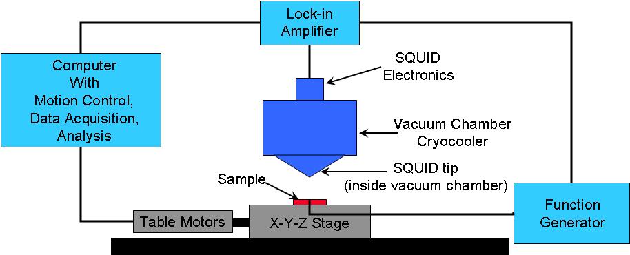
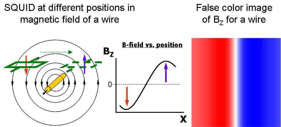
When an electrical
current is passed through a wire, the wire produces
a magnetic field. The SQUID can be used to
measure the Z component of the magnetic field from
current carrying wires, and a magnetic image can be
generated.
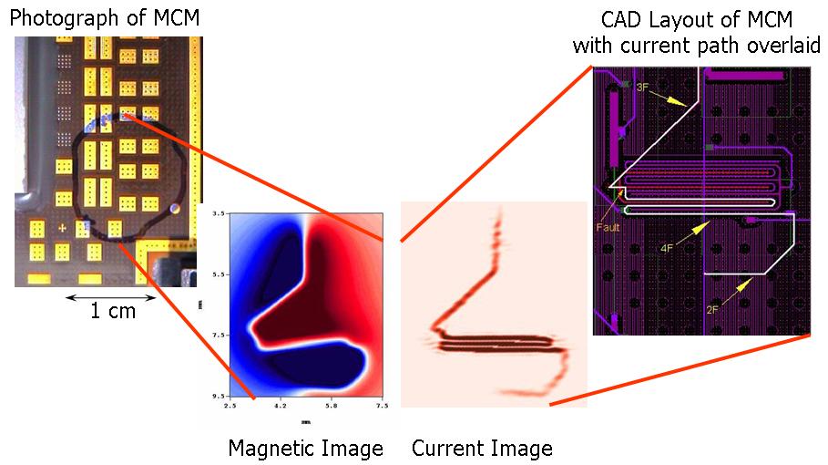
A magnetic inversion
algorithm can then be used to calculate a map of the
currents that produced the magnetic field.
This allows the SSM to "see through" solid material
and determine the location of defects (short
circuits) in circuits. Below is an example of
images produced by a defect that was located inside
a Multi-Chip Module (MCM). (images courtesy of
Neocera, LLC)
The SQUID microscope
can also be used to image static magnetic fields
such as those produced by the magnetic ink in a
dollar bill or even a laser-printed Trevecca logo.
This false-color image is from a Trevecca logo that
was printed out, magnetized, then scanned with the
SQUID microscope.

|